What is Dental Radiology?
Dental radiology is an imaging method used to detect the discomfort in oral and dental disorders, and to determine the treatment process and method. With the radiography imaging method, it helps to easily detect caries, inflammation, disorders in the bones or problems in the tooth roots that cannot be seen during the examination.
With the dental radiology method, we can detect diseases before they progress and find solutions in the short term. Radiography imaging method is a very short and comfortable procedure. In our clinic, any situation that may disturb our patients is done before they develop.
Is Dental Radiology Harmful?
Since the method we use is a radiography imaging method, there is definitely an X-ray that passes from the devices used to the patient. As in every field, technology has developed a lot in the field of health, and the X-ray that the patient receives during imaging is almost the same as the X-ray received while watching television or using the phone. Dental radiology is used in children as in adults. The harm is minimal as in adults. There is no harm for children. dental radiology;
- Detection of joint disorders,
- Detection of cysts in teeth and bones,
- Detection of invisible and root bruises,
- Detection of fractures and dislocations in the tooth and jaw,
- Detection of salivary gland diseases,
- Appearance of the condition of the tooth for surgical intervention
- The canal serves and functions in many areas such as patient follow-up before and after treatment.
As a result of the examination of our doctor, there are types of x-rays that the patient wants according to his condition. These types of x-rays;
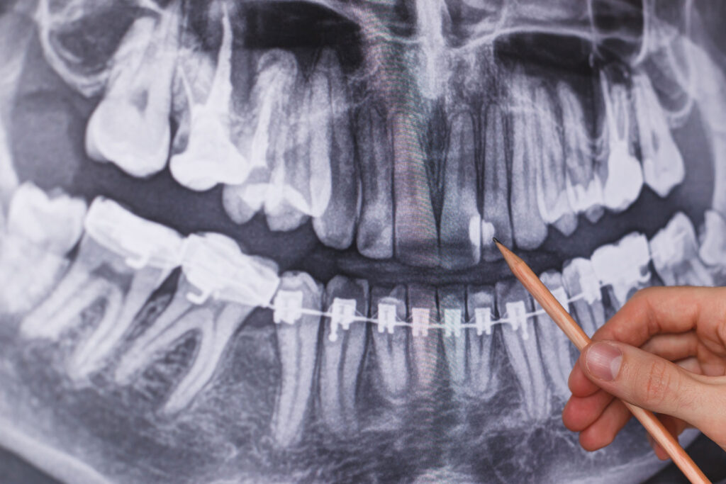
- Panoramic radiography showing the structure and condition of all teeth and bones in the mouth,
- Periapical radiograph showing two or three teeth together that the patient is complaining about or that the doctor wants to see more closely,
- Bitewing radiograph showing the right or left segment of the mouth used to detect caries,
It is in the form of three-dimensional imaging methods (Dental Volumetric Radiography-DVT) that can be taken before surgical procedures such as implant planning.
How Often Can Radiography Be Taken?
The frequency of radiographs varies depending on the discomfort and process determined as a result of the doctor’s examination of the patient. The duration is determined according to the patient’s medical history. For some patients, our doctors consider 2-year peridots sufficient.
State-of-the-art radiography instruments are used in our clinic. We perform imaging in a practical way for our pediatric and adult patients without any discomfort. Imaging methods have an important place in the detection and treatment of diseases in oral health. Our clinic is among the clinics that use this method most effectively.
Have you seen our orthodontic service?